Sunday, 20 December 2020

Endocrine System 1


The Hypothalamus

The hypothalamus is the basal part of diencephalon, forebrain.

It contains several groups of neurosecretory cells called nuclei which produce hormones. These hormones regulate the synthesis and secretion of pituitary hormones. However, the hormones produced by hypothalamus are of two types, the releasing hormones (which stimulate secretion of pituitary hormones) and the inhibiting hormones (which inhibit secretions of pituitary hormones).

Hypothalamic hormone called Gonadotrophin releasing hormone (GnRH) stimulates the pituitary synthesis and release of gonadotrophins.

Somatostatin from the hypothalamus inhibits the release of growth hormone from the pituitary.

Hormones originating in the hypothlamic neurons, pass through axons, and are released from their nerve endings.

Hormones reach the pituitary gland through a portal circulatory system and regulate the functions of the anterior pituitary.

The posterior pituitary is under the direct neural regulation of the hypothalamus.
 

The Pituitary Gland

The pituitary gland is located in a bony cavity called sellatursica and is attached to hypothalamus by a stalk. It is divided anatomically into an adenohypophysis and a neurohypophysis. 

Location of Pituitary Gland

The pars distalis region of pituitary, commonly called anterior pituitary, produces growth hormone (GH), prolactin (PRL), thyroid stimulating hormone (TSH), adrenocorticotrophic hormone (ACTH), luteinizing hormone (LH) and follicle stimulating hormone (FSH).

Pars intermedia secretes only one hormone called melanocyte stimulating hormone (MSH).

In humans, the pars intermedia is almost merged with parsdistalis.

Neurohypophysis (pars nervosa) also known as posterior pituitary, stores and releases two hormones called oxytocin and vasopressin, which are actually synthesised by the hypothalamus and are transported axonally to neurohypophysis.

Over-secretion of GH stimulates abnormal growth of the body leading to gigantism and low secretion of GH results in stunted growth resulting in pituitary dwarfism.

Prolactin regulates the growth of the mammary glands and formation of milk in them.

TSH stimulates the synthesis and secretion of thyroidhormones from the thyroid gland. ACTH stimulates the synthesis and secretion of steroid hormones called glucocorticoids from the adrenal cortex. LH and FSH stimulate gonadal activity and hence are called gonadotrophins. In males, LH stimulates the synthesis and secretion of hormones called androgens from testis. In males, FSH and androgens regulate spermatogenesis. In females, LH induces ovulation of fully mature follicles (graafian follicles) and maintains the corpus luteum, formed from the remnants of the graafian follicles after ovulation. FSH stimulates growth and development of the ovarian follicles in females.

MSH acts on the melanocytes (melanin containing cells) and regulates pigmentation of the skin.

Oxytocin acts on the smooth muscles of our body and stimulates their contraction. In females, it stimulates a vigorous contraction of uterus at the time of child birth, and milk ejection from the mammary gland.

Vasopressin acts mainly at the kidney and stimulates resorption of water and electrolytes by the distal tubules and thereby reduces loss of water through urine (diuresis). Hence, it is also called as anti-diuretic hormone (ADH).

Blood supply to pituitary or Hypophyseal portal system: A pair of posterior hypophysial arteries and a pair of anterior hypophysial arteries provide blood to the pituitary gland. Posterior arteries supply blood to the pars nervosa, and anterior arteries supply blood to the hypothalamus and pars distalis. Adenohypophysis has dual blood supply by means of a "circle of willis". The anterior hypophyseal artery which bring blood into this circle bifurcates in to two branches outside the lobe. One branch supplies the adenohypophysis and other supplies the hypothalamus. The veins that dratn the blood from hypothalamus, run into the pars distalis through pars tuberalis and divide into capillaries. Those veins are therefore, called portal hypophyseal veins. These constitute a hypothalamo hypophyseal portal system. Hypothalamic hormone reaches anterior pituitary by portal system. 

Blood Supply to Pituitary

Nanism or ateliosis: Hyposecretion (undersecretion) of growth hormone is childhood results into a blunted growth of body. Growth of all organs is retarded. Growth of bones at their epiphysial ends stops. Hence, the bones do not grow in length, so that the body remains a dwarf. This pituitary dwarfism is called nanism or ateliosis.

Midgets: Unlike the thyroid cretinism, the development of brain is normal in pituitary dwarfs, but like thyroid cretinism, the pituitary dwarfs are also infertile. The dwarfs of circuses are pituitary dwarfs. These are called midgets.

Pituitary myxoedema: Undersecretion of growth hormone during adolescence (between 13 to 22 years of age) restricts body height, so that the person remains short-statured. Under secretion after growth period (about the age of 22) causes pituitary myxoedema whose symptoms are almost similar to those of thyroid myxoedema. These include old age symptoms, such as reduced BMR and protein synthesis, graying and falling of hair, dryness of skin, reduced BP and low body temperature, insomnia, and weakness of muscles, vision and wisdom. Due to accumulation of mucus under the skin, the body becomes puffy, but weak. Genitalia weaken, causing sexual disability. Hence, the person becomes disheartened.

Proportionate gigantism: Hypersecretion (oversecretion of growth hormone during growth period (childhood and adolescence) causes excessive growth (hyper growth) of all body parts, resulting into a symmetrically giant body. This is called proportionate gigantism.

Disproportionate gigantism or acromegaly: The concerned person may attain a height of 8 feet or ever more. Oversecretion of growth hormone after growth period also causes gigantism, but in this the long bones do not grow in length due to dosed epiphyseal plate at their ends, but the bones of hands, feet, lower jaw and rib cage thicken. Simultaneously, eyelids, lips, tongue, nose, chin, etc also enlarge. Soles, palms and forehead become wrinkled. Skin thickens and becomes wrinkled. Skin thickens and becomes coarse and fluffy (hirsutism), Consequently, the body becomes ugly like a gorilla. This is called disproportionate gigantism or acromegaly. It is common in men and rare in women.

Kyphosis: In some cases, the backbone bends and thickens, causing hunchback condition (kyphosis). Breasts enlarge and mammary gland may yield milk. The patients often complain of headache, sexual disorders, muscular pain, and impaired vision and memory.

Hypersecretion of growth hormone raises blood glucose level (hyperglycemia) which may cause diabetes mellitus.

Ketosis: Increased breakdown of fat may release ketone bodies, mainly acetoacetic acid, in blood, causing ketosis.

Prolactin hormone is secreted both in males as well females. In males it influence sexual behavior. Its hypersecretion may hinder menstruation.

(ii) May cause impotency

(iii) In pigeons and doves, it stimulates the epithelia cells of crop in both males and females to secrete "pigeon milk" for nutrition of newly hatched infants.

The pituitary gland plays most important regulator; role in the body. Besides regulating growth, sex and general behaviour it also regulates the secretory activities of other principal endocrine glands and cells. Most appropriately, therefore pituitary has been referred to as "The Master Gland' of body, or the "Chief Executive of Endocrine System", or "The Leader of Endocrine Orchestra" 

Hormones of Adenohypophysis

The Pineal Gland

The pineal gland is located on the dorsal sir forebrain. Pineal secretes a hormone ca melatonin.

Melatonin plays a very important role ir regulation of a 24-hour (diurnal) rhythm of body. For example, it helps in maintaining the normal rhythms of sleep-wake cycle, body temperature. In addition, melatonin also influence metabolism, pigmentation, the menstrual cycle well as our defense capability.

Light falling on the retina of the eye decreases melatonin production, darkness stimulates melatonin synthesis. Girls blind from birth attain puberty earlier than normal, apparently because there is no inhibitory effect of melatonin on ovarian function.

Serotonin, a neurotransmitter found in other locations in the brain, is also found in the pineal gland. Research evidence is accumulating to support the idea that the pineal gland may be involved in regulating cyclic phenoinena in the body. Melatonin also is a potent antioxidant. Melatonin causes atrophy of gonads in several animals.


Thyroid Gland

The thyroid gland is composed of two lobes which are located on either side of the trachea. Both the lobes are interconnected with a thin flap of connective tissue called isthmus. 

(a) Thyroid Gland (b) Follicles Suspended in Stroma of a Lobule

The thyroid gland is composed of follicles and stromal tissues. Each thyroid follicle is composed of follicular cells, enclosing a cavity. These follicular cells synthesise two hormones, tetraiodothyronine or thyroxine (T4) and triiodothyronine (T3).

Iodine is essential for the normal rate of hormone synthesis in the thyroid. Deficiency of iodine in our diet results in hypothyroidism and enlargement of the thyroid gland, commonly called goitre.

For secreting the iodinated hormones in normal amounts, the thyroid daily utilizes about 150 micrograms (0.15 milligram) of iodine. Obviously, a person must daily obtain 150mg of iodine from food. We can obtain this from dairy products, drinking water, seafood, etc. If obtained more than this, we excrete the excess iodine in urine.

Iodine of food is absorbed and circulated in blood in the form of iodide ions (I-). Follicular cells of thyroid very actively obtain these ions from blood by active transport. That is why, the concentration of I- in these cells normally remains about 50 to 250 times more than in blood. These cells possess peroxidase enzyme in abundance. Peroxidase continuosly oxidizes iodide ions into molecular iodine (2I- ® I2), Iodine is, then, released by follicular cells into follicular' cavity.

Thyroid hormones play an important role in the regulation of the basal metabolic rate. These hormones also support the process of red- blood cell formation. Thyroid hormones control the metabolism of carbohydrates, proteins and fats, Maintenance of water and electrolyte balance is also influenced by thyroid hormones.

Thyroid gland also secretes a protein hormone called thyrocalcitonin (TCT) which regulates the blood calcium levels.

Cretinism: It is disease of infants, called cretins. It is characterised by Decreased BMR (50% than normal); stunted growth; retarded mental development so low I.Q., delayed puberty; decreased body temperature, heart rate, pulse rate, blood pressure and cardiac output; reduced urine output; decreased sugar level in blood, pigeon's chest (chest bulging forward in sternal region). Cretinism can be congenital (absence of thyroid due to genetic defect) or endemic (absence of iodine in diet). It can be corrected by thyroxin administration.

Myxoedenta: It occurs due to deficiency of thyroxine in adults like cretinism, it also has low (BMR) (by 30 - 40%); low body temperature, reduced heart rate, pulse rate, blood pressure and cardiac output, low sugar and iodine level in blood etc. But the peculiar feature of myxoedema is that face and hands become swollen due to deposition of albuminous myxomatous tissue. It can also be corrected by thyroxine administration.  

Endemic or simple goitre or colloid goitre: It occurs due to deficiency of iodine in drinking water. It is non-genetic (sporadic goitre is a genetic disease). It is characterized by enlargement of thyroid gland due to increase in number and size of acinal cells of thyroid gland. It is more common in people of hilly region. To prevent goitre, the table salt is being iodised these days.

Hashimoto's disease: It is called auto-immune thyroiditis and occurs due to age factor, injury-surgery, wrong treatment or injection thyroid gland causing hyposecretion of thyroxine. When thyroxine secretion falls upto minimal limit, the antibodies are formed which destroy the thyroid gland.

Simple goitre: Hyperthyroidism may be simply because of overactive cells of a normal gland, or because of an enlargement of the gland, causing goitre.

Exophthalmic goitre : Such a goitre is called exophthalmic goitre, because it is usually accompanied with some asymmetrical protrusion (Exophthalmos) of the eyeballs, imparting an angry, frightened, or staring look to the patient, Protrusion of eyeballs is due to accumulation of mucus in eye orbits.

Grave's or Basedow's disease: Enlargement of the gland is usually due to a diffused growth.

Plummer's disease or Toxic Adenoma: It is due to formation of one or more hypersecretory nodules plummer's diseases or Toxic Adenoma in the gland. 


Parathyroid Gland

In humans, four parathyroid glands are present on the back side of the thyroid gland, one pair each in the two lobes of the thyroid gland. The parathyroid glands secrete a peptide hormone called parathyroid hormone (PTH). The secretion of PTH is regulated by the circulating levels of calcium ions.

(a) Parathyroid Gland Embedded in the Surface of Thyroid (b) Ultrastructure of a Parathyroid

Parathyroid hormone (PTH) increases the Ca2+ levels in the blood. PTH acts on bones and stimulates the process of bone resorption (dissolution/ demineralisation). PTH also stimulates reabsorption of Ca2+ by the renal tubules and increases Ca2+ absorption from the digested food. It is, thus, clear that PTH is a hypercalcemic hormone, i.e., it increases the blood Ca2+ levels. Along with TCT, it plays a significant role in calcium balance in the body.

Hypoparathyroidism (Hyposecretionof parathormone)

(i) It is rare, however, in undersecretion of parathormone, the level of calcium in ECF falls (hypocalcemia), and that of phosphates rises (hyperphosphatemia). This causes neuromuscular hyperexcitability, excessive perspiration gooseflesh (raising of hairs and prickly sensation in skin), cooling of hands and feet, painful muscular spasms and convulsions, and trembling.

(ii) Sometimes some skeletal muscles, usually of hands and feet, fail to relax after a contraction and remain in "sustained contraction". This is called "Tatany”. Tetany of larygncal, thoracic, and phrenic muscles, which help in breathing, causes death, because the patient fails to breathe (asphyxia). (iii) Childhood hypoparthyroidism retards growth, particularly of bones, teeth, hair and brain. Vitamin D is administered to such children.

Hyperparathyroidism (Hypersecretion of parathonnone)

(i)  Osteoporosis: Oversecretion of parathormone is rare and occurs usually due to overgrowth of one or more parathyroid glands. It causes demineralization of bones which, therefore, become soft, weak, distorted and fragile. This is called osteoporosis.

(ii) Hypercalcemia: Simultaneously, due to a sharp rise in calcium level in blood and ECF (hypercalcemia) and a sharp fall in phosphate level (hypophosphatemia), muscles and nerves are weakened.

(iii) Hypercalciurea: Calcium is excreted in urine (hypercalciurea), thirst increases owing to copius urination, appetite is lost, constipation and headache become common, and often, kidney stones are formed. The only treatment so far known is removal of extra part of the glands by operation. 
 

Thymus

The thymus gland is a lobular structure located on the dorsal side of the heart and the aorta. 

Location of Thymus Gland

This gland secretes the peptide hurmones called thymosins. Thymosins playa major role in the differentiation of T lymphocytes, which provide cell-mediated immunity. In addition, thymosins also promote production of antibodies to provide humoral immunity. 

Thymus is degenerated in Old individuals resulting in a decreased production of thymosins. As a result, the immune responses of old persons become weak. 
 

Adrenal Gland

Our body has one pair of adrenal glands, one at the anterior part of each kidney.

 Adrenal Gland With a Part Cut to Show Cortex and Medulla

The gland is composed of two types of tissues. The centrally located tissue is called the adrenal medulla, and outside this lies the adrenal cortex.

The adrenal medulla secretes two hormones called adrenaline or epinephrine and noradrenaline or norepinephrine. These are commonly called as catecholamines. Adrenaline and nor adrenaline are rapidly secreted in response to stress of any kind and during emergency situations and are called emergency hormones or hormones of Fight or Flight. These hormones increase alertness, pupilary dilation, piloerection (raising of hairs), sweating etc. Both the hormones increase the heart beat, the strength of heart contraction and the rate of respiration. Catecholamines also stimulate the breakdown of glycogen resulting in an increased concentration of glucose in blood. In addition, they also stimulate the breakdown of lipids and proteins.

The adrenal cortex can be divided into three layers, called zona reticularis (inner layer), zona fasciculata (middle layer) and zona glomerulosa (outer layer). 

 Adrenal Gland With a Part Cut to Show Cortex and Medulla

The adrenal cortex secretes many" hormones, commonly called as corticoids. The corticoids, which are involved in carbohydrate metabolism are called glucocorticoids. In our body, cortisol is the main glucocorticoid. Corticoids, which regulate the balance of water and electrolytes in our body are called mineralocorticoids. Aldosterone is the main mineraiocorticoid in our body.

Glucocorticoids stimulate, gluconeogenesis, lipolysis and proteolysis; and inhibit cellular uptake and utilisation of amino acids. Cortisol is also involved in maintaining the cardio-vascular system as well as the kidney functions. Clucocorticoids, particularly cortisol, produces antiinflamatory reactions and suppresses the immune response. Cortisol stimulates the RBC production. Aldosterone acts mainly at the. renal tubules and stimulates the reabsorption of Na+ and water and excretion of K+ and phosphate ions. Thus, aldosterone helps in the maintenance of electrolytes, body fluid volume, osmotic pressure and blood pressure.

Small amounts of androgenic steroids are also secreted by the adrenal cortex which play a role in 'the growth of axial hair, pubic hair and facial hair during puberty.

Hyposecretion: This may be a genetic defect.

Undersecretion of adrenocorticoids (hypocorticism) causes Addison's disease which is relatively rare and occurs in both men and women between the ages of 20 to 40 years. This disease was first described by Thomas Addison in 1849, 1855. It is maintained in following symptoms:

Owing to low aldosterone and gluco-corticoides level in blood, considerable amount of sodium ions an water is excreted in urine, leading to dehydration, low blood pressure, and weakness, all symptoms of a peculiar, Addinosonean anaemia which is different from common pernicious anaemia resulting from entirely different causes like diarrhoea, cholera, etc.

Owing to low cortisol level, glucose level also falls in blood (hypoglycemia). This sharply reduces BMR in body cells. Due to hypoglycemia and hyperkalemia (increased K+ level in blood) efficiency of brain, liver, skeletal and cardiac muscles, etc declines, Body temperature also falls. Hearibeat may even stop, causing death.

Decreased cortisol level induces gastro intestinal disorders, resulting in loss of appetite, nausea, vomiting, diarrhoea, abdominal pain and restlessness.

Due to a sharp decline in body's chemical defense and resistance, sensitivity to cold, heat, infection, poisoning and other adverse condition increases. Acute hypocorticism is catastrophic and reentrance sensitivity to cold, heat, infection, poisoning and other adverse conditions increases. Acute hypocorticism is catastrophic and threatens life. Complete destruction of removal of adrenals causes death in' a short time, principally because of loss of excessive sodium in urine.

Addison's disease also causes an increase in the number of WBCs, resulting into eosinophilia, lymphocytosis, leucocytosis, etc.

Uridersecretion of sex hormones causes impotence in males and disorders of menstrual cycle in females.

Excessive deposists of melanin, particularly in the skin of open parts of body like face, hands, feet neck, teats, etc cause deep bronzing of skin in these part.

An increase in H+ concentration in blood may cause acidosis.

Hypersecretion: Oversecretion of adrenocorticoids (hypercorticism) causes following disorders and disease.

Glucose level rises in blood (hyperglycemia). This may lead to diabetes mellitus.

Irregular deposits of fat, particularly in thoracic parts and face, imparts asymmetrical shape to the body. The face becomes red and rounded (moon face), shoulders swell (buffalo humps) and abdomen dilates and often shows lines of stretching: All these are symptoms of Cushing's 'disease (Cushing, 1932). Patients may die from brain haemorrhage, cardiac arrest, pneumonia, etc.

Retention of sodium and water in the ECF increases blood pressure, causing severe hypertension and associated symptoms like severe headache.

Excessive loss of potassium in urine causes potassium deficiency (hypokalemia). This leads to muscular weakness and convulsions and' nervous disorders, and may even cause tetany and paralysis, copious and frequent urination (polyuria) and thirst, bed urination (nocturia), etc. Similarly, excessive loss of H+ in urine may cause alkalosis.

Excessive mobilization of materials from all parts of body had widespread deteriorating effects. For instance, mobilization of proteins from all cells causes (issue wasting. Similarly, mobilization from bones renders the bones weak and fragile (osteoporosis). 

Neural control Nerve conduction.


Neural Control and Coordination


Main topics at Glance-


The Nervous System is a very important part of the animal body that coordinates voluntary and involuntary actions of the body.

What is Nerve Impulse?

Any stimulus or signal that activate or inhibit any gland, muscle, organ, tissue or any nerve cell as known as Nerve impulse. For Example: When we touch a hot plate, a signal is transmitted to brain due to which we respond by removing our hand.

There are two  types of Nervous System in vertebrates:

  • Central Nervous System

  • Peripheral Nervous System

Divisions of Nervous System

Fig.1. Divisions of Nervous System

What is Central Nervous System?

The central Nervous System comprises of Brain and the Spinal Cord.
 

Explain the Structure of Human Brain.

The Human Brain is covered by a membrane known as Meninges. Meninges are three in number. The Outermost DuramaterMiddle Arachnoid and Innermost Piamater.

The longitudinal fissure divides the brain into two hemispheres known as Right Cerebral Hemisphere and Left Cerebral Hemisphere.

The Brain is divided into three parts: ForebrainMidbrain and Hindbrain.
 

Forebrain

The Forebrain comprises of Cerebrum,Thalamus and Hypothalamus.

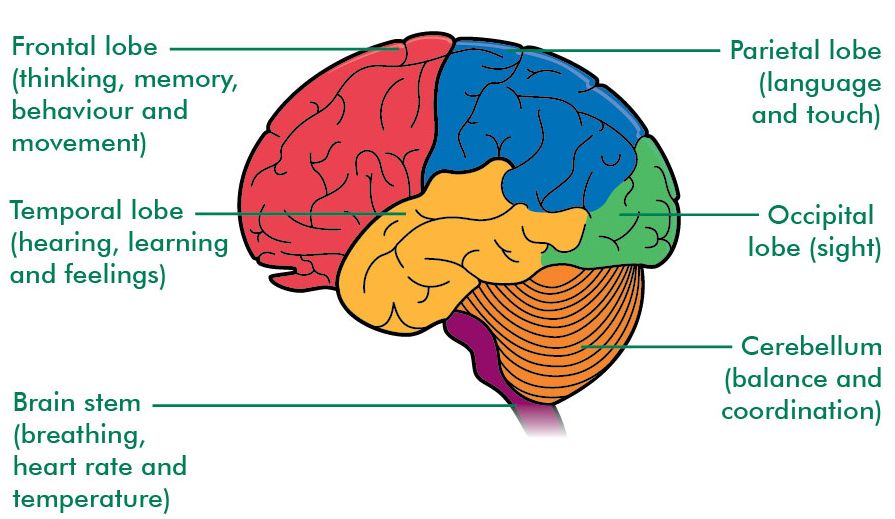
  • Cerebrum is the largest part of the human brain. It is divided into four lobes: Occipital LobeParietal LobeTemporal Lobe and Frontal Lobe.

Different Lobes of Cerebrum and their function

Fig.2. Different Lobes of Cerebrum and their function

Thalamus is olive shaped structure which performs sensory functions. It directs sensory input from the eyes, ears, tongue and from other sense organs.

Hypothalamus is located below the thalamus as the name suggests. It is important in monitoring hormone concentrations, water concentrations, temperature of the body etc.
 

Midbrain

It is a small part of the brain. It is associated with Vison, Hearing, Sleep, Arousal, Temperature etc.
 

Hindbrain

It consists of Cerebellum, Pons and Medulla Oblongata.

  • Cerebellum is involved in controlling attention, language, equilibrium, posture, etc.

  • Pons controls digestive and respiratory movements.

  • Medulla Oblongata connects skull with the spinal cord. It helps in controlling Blood Pressure, Heart Rate, Respiratory Movement, Vomiting etc.
     

What are Neurons/Nerve Cell?

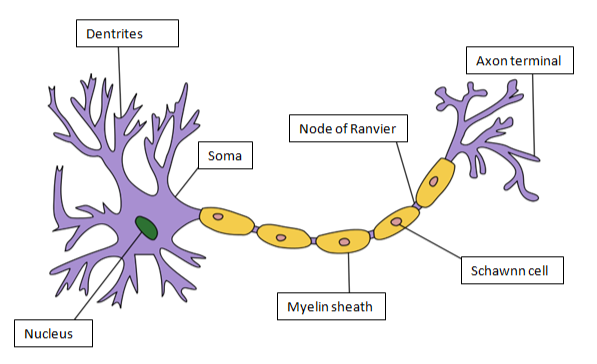
The structural and the functional unit of brain is NeuronNeuron is divided into Cell BodyAxon and Dendrites. Cell body/soma of the neuron contain nucleus. It also contains Nissl Granules, composed of endoplasmic reticulum. Dendrites are branched projections from the cell body. Axon is a long, slender in shape that transmits nerve impulse from one neuron to another. 
Structure of Neuron

Fig.3. Structure of Neuron

The cells found in axon are known as Schawnn Cell. Myelin sheath is schawnncells. Myelin sheath is an electrically insulating layer which is required for fast and efficient nerve impulse transmission. A gap between adjacent Schawnn Cells is known as Nodes of Ranvier.


Example of different types of Neurons

  • Sensory Neurons transmit nerve impulses from sensory receptors to central nervous system.

  • Motor Neurons transmit nerve impulses from central nervous system to muscle or a gland.

  • Inter Neurons connects different neurons within the brain and spinal cord.

Spinal Nerves
Spinal Cord

It is a thin, tube-like structure which extends from the medulla of the brain to the lumbar region of vertebral column. It helps in transmission of nerve impulses from the brain to the rest of the body. It also coordinates reflex action.

There are 31 pairs of Spinal Nerves on each side of the vertebral column. These Spinal Nerves are grouped into:

  • 8 pairs of cervical nerves

  • 12 pairs of thoracic nerves

  • 5 pairs of lumbar nerves

  • 5 pairs of sacral nerves

  • One pair of coccygeal nerves

The Spinal Cord is made up of White Matter and Gray Matter. The butterfly shaped structure present at the center of the spinal cord is known as Gray Matter

Structure of Spinal Cord

Fig.5. Structure of Spinal Cord

The region around the gray matter is known as White Matter. White matter contains nerves that are covered with Myelin Sheath whereas gray matter contains nerves without myelin sheath.
 

What is Reflex Action?

The involuntary response towards any stimulus is known as Reflex Action.

What is Reflex Arc?

The pathway of reflex action is known as Reflex Arc. Some sensory neurons do not pass directly into the brain; they innervate in the spinal cord. This reduces the time of reflex action.

What is Peripheral Nervous System?

It consists of nerves and ganglia. Ganglia is a cluster of nerves. Peripheral Nervous System connects central nervous system to different organs and limbs.

Peripheral nervous system is divided into Autonomic Nervous System and Somatic Nervous System.  Autonomic nervous system is associated with communication to internal organs and glands. Somatic nervous system communicates with sensory organs and voluntary muscles.

Autonomic Nervous System is further divided into Parasympathetic Nervous System and Sympathetic Nervous System.


Difference between Sympathetic and Parasympathetic Nervous System


Sympathetic Nervous System

Parasympathetic Nervous System

Type of Control

Involuntary

Involuntary

Number of neurons per message

Two (Preganglionic shorter than Postganglionic)

Two (Preganglionic longer than Postganglionic)

Location of motor fiber

Thoracolumbar spinal nerves

Cranial (e.g., vagus) and sacral spinal nerves

Neurotransmitter

Norepinephrine

Acetylcholine

Effectors

Smooth and cardiac muscle, glands

Smooth and cardiac muscle, glands


Human Sense Organs

Humans have five sense organs, They are: Eyes, Ears, Nose, Skin and Tongue.

The structure of Eye and Ear is explained below:
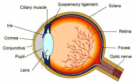
 

Structure of Human EyeStructure of the Eye

Eye is a sense organ for sight. It consists of three layers: Outermost ScleraMiddle Choroid which is supplied with blood vessels and Inner Retina. The membrane that covers the sclera is known as Conjunctiva. Retina contains light sensitive cells known as Rods and Cones. Rods are cells that allow vision in dim light whereas cones help in color vision. Light enters the eye via cornea. 

The nerve that connect eye to brain is known as Optic Nerve. Transparent lens focuses light on retina which helps in vision. The muscles that contracts and relaxes the lens is known as Suspensory Ligament. This is dependent on the amount of light that falls on the lens.

The colored part of the eye that controls the amount of light that enters the eye is known as Pupil. Fovea is an exact location where light is focused on the retina. 
 

Structure of Ear

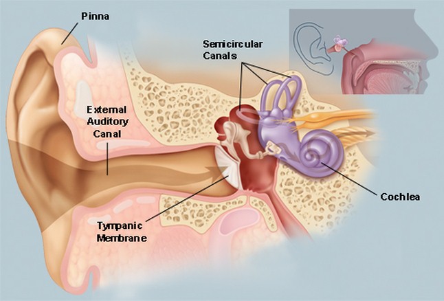
Ear is meant for hearing. The ear is divided into External EarMiddle Ear and Inner Ear. The outer part of the ear is known as Ear Pinna. Ear pinna is covered by a membrane known as Tympanic Membrane

Structure of the Ear

Fig. 7. Structure of the Ear

When sound strikes the pinna it vibrates and vibrations are passed to cochlea which is a part of the inner ear. There are fluid-filled canals present that are attached to cochlea, are known as Semi-Circular Canals. These canals help in maintaining equilibrium or balance of the body.

Stimulus and Response

It is defined as any detectable change in the internal or the external environment. The ability of an organism to react against the stimulus is known as Response.


Synapse

The junction between the two Nerve cells is known as Synapse.
 

Second Brain in Humans

The Enteric Nervous System is known as Second Brain in Humans.
 

Saltatory Conduction

During Nerve Impulse transmission, the nerve impulse jumps over the Nodes of Ranvier. This mode of nerve impulse transmission is known as Salutatory Conduction. This is the fast mode of nerve impulse transmission. 

MCQ MOLECULAR BASIS OF INHERITANCE

  1. Amino acid sequence, in protein synthesis is decided by the sequence of ( a ) r RNA ( b ) t RNA ( c ) m RNA ( d ) c DNA 2. Antipa...