Tuesday, 19 September 2023

BODY FLUID AND CIRCULATION XI


Body Fluids and Circulation

Table of Content

Blood

It is a fluid connective tissue consists of matrix, plasma and formed elements.
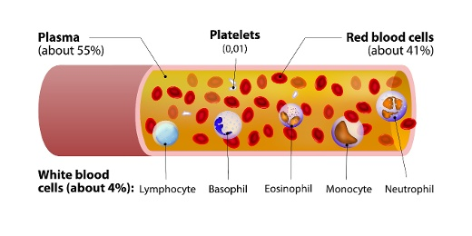
Fig. 1. Elements of the blood

Plasma

It is a straw colored, viscous fluid which constitutes about 55% of the blood plasma. The major proteins found in plasma are fibrinogen, globulins, and albumins. Fibrinogens functions during blood clotting. Albumins maintain osmotic balance in the body. Globulins are defensive in nature. Minerals such as Sodium IonsCalcium IonsMagnesium IonsBicarbonate Ions. Apart from this amino acid, glucose is also present in plasma.

Formed Elements

Formed Elements include erythrocytes, leucocytes, and blood platelets.
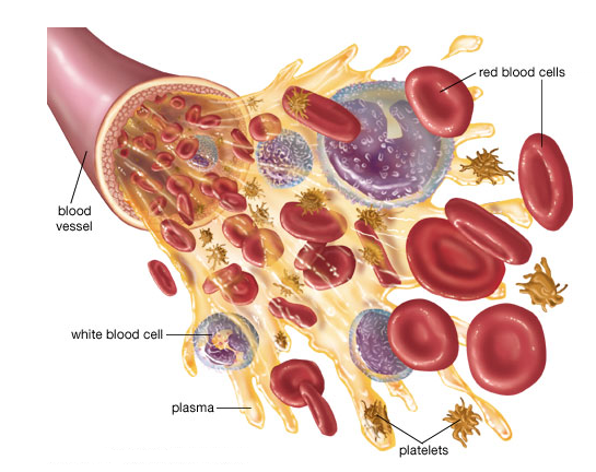
Fig. 2. Formed elements in blood

Erythrocytes also known as Red Blood Cells. They are the most abundant cells in the blood. They are formed in red bone marrow of adults. They are biconcave in shape and enucleated (without the nucleus). They carry iron containing protein known as Hemoglobin. Hemoglobin helps in the transport of oxygen and carbon-dioxide in blood. The average life span of RBCs is 120 days.

Leucocytes also known as White Blood Cells. They are nucleated cells which do not contain hemoglobin, so they appear colorless. They are of two types- granulocytes and agranulocytes.

Neutrophils, Basophils and Eosinophils are granulocytes. Lymphocytes and Monocytes are agranulocytes.

Neutrophils are known as Polymorphonuclear Leucocytes. Out of all the three granulocytes, neutrophils are most abundant. They are phagocytic cells.

Basophils are least in number in comparison to other granulocytes. They secrete serotonin, histamine, and basophils. So, basophils are involved in inflammatory reactions.

Eosinophils are involved in allergic reactions.


Fig. 3. White blood cells and platelets

Blood platelets are also known as Thrombocytes. They are involved in blood clotting. Decrease in the number of blood platelets can lead to loss of blood in the body.

Blood Groups

Blood grouping is divided into- ABO blood group and Rh group.

ABO blood grouping

ABO blood grouping is done based the presence or absence of certain antigens on the surface of RBCs. The antigen present on the surface of RBCs can be A or B. There are 4 types of blood group are A, B, AB and O group.

Fig. 4. Blood groups and antigen on red blood cells

The above table depicts blood groups and donor compatibility. As O blood group do not have any antigen on their surface, they are said to be universal donor whereas AB are considered universal recipients as they contain both the antigen of their surface. Blood transfusion is done only based on blood group of the donor and recipients.

Rh grouping

Rh is also an antigen like rhesus monkeys. Individuals having Rh antigen on RBCs are considered as Rh positive. Those which are without Rh antigen are considered Rh negative. If Rh -ve person receives Rh +ve blood, the Rh -ve individual will start producing antibodies against it. So, Rh group should also be tested at the time of blood transfusion.

An important case of Rh mismatching has been observed between the Rh -ve blood of a pregnant mother with Rh +ve blood of the fetus. Rh antigens of the fetus do not get exposed to the Rh-ve blood of the mother in the first pregnancy, due to placenta. But during the delivery of the first child, there is a possibility of mixing of the blood of mother with child.

In such cases, mother starts to produce antibodies against the Rh antigen. So, in the next pregnancy, the Rh antibodies from the Rh -ve mother can leak into the blood of the Rh +ve fetus and can destroy the fetal RBCs. This leads to agglutination of red blood cells. This condition is known as Erythroblastosis Foetalis. Fetus will anemic and suffers from jaundice. This can be avoided by injecting anti-Rh antibodies to the mother instantly after the delivery of the first child.

Coagulation of Blood

Blood coagulation is also known as Blood Clotting. Blood responds by clotting or coagulation after any injury or trauma. This helps in preventing excess loss of blood. When we get injured, after some time reddish brown scum is formed at the site of injury. This is known as clot which is made up of network of threads known as Fibrils. This network contains dead and damaged formed elements of blood. Fibrils are formed from the conversion of inactive fibrinogen in presence of enzyme thrombin. Thrombin is also formed from inactive form known as ProthrombinThrombokinase is required for the conversion of prothrombin into thrombin.

Platelets release certain factors to begin blood clotting. Calcium ions play an important role during Blood coagulation/clotting.
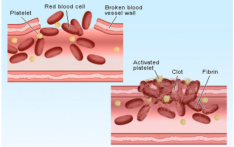
Fig. 5. Blood coagulation

Lymph

Blood circulates in blood capillaries in tissues. Some water along with some water-soluble substances leak out and enter the interstitial space. This is known as Tissue Fluid or Interstitial Fluid. Exchange of gases, nutrients between the blood and the cells occurs through this fluid.

A network of vessels that collects this fluid and drain it to major veins is known as Lymphatic System. Lymphatic system contains a fluid known as Lymph. Lymphocytes which are an important cell of the immune system are present in lymph.
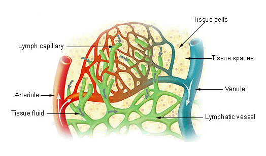
Fig. 6. Lymph

Circulatory Pathways

There are two types of circulatory system- Open Circulatory System and Closed Circulatory System.

When blood flow in open spaces known as Lacunae and Sinuses, it is known as Open Circulatory System it is present in molluscs, arthropods, etc.

When blood flow in closed vessels it is known as Closed Circulatory SystemFor Example, humans. Closed circulatory possess an advantage over open circulatory system, as blood can flow in a regulated manner in case of closed circulatory system.

Vertebrates have muscular, pumping organ known as Heart. Fishes have 2 chambered heart. Amphibians have 3 chambered heart except crocodiles (4 chambered heart). Birds, reptiles, and humans have 4 chambered heart.

Human Circulatory System

It includes heart, vessels, and blood. Heart is mesodermal in nature. It is located in the thoracic cavity, in between the two lungs.

The double membrane that surrounds the heart is known as Pericardium. Pericardium encloses the pericardial fluid. Heart is 4 chambers with 2 atria and 2 ventricles. A thin wall separates the left and the right atria is known as Intra-atrial Septum whereas left and right ventricle are separated by thick intra-ventricular septum.
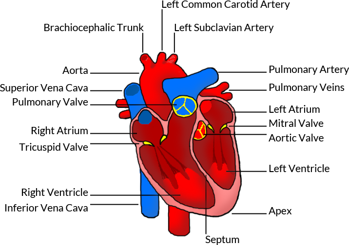
Fig. 7. Human heart

The opening between the right atrium and the right ventricle is guarded by a valve known as Tricuspid Valve, whereas a bicuspid or mitral valve guards the opening between the left atrium and the left ventricle. The openings of the right and the left ventricles into the pulmonary artery and the aorta respectively are provided with the semilunar valves. Valves in the heart allows the blood to flow in one direction and thus preventing backflow of blood.

Heart is a muscular organ and the muscles of the heart are known as cardiac muscles. A specialized cardiac musculature called the nodal tissue is also dispersed in the heart. One which is present in the upper right corner of the right atrium is known as Sinoatrial Node or SA Node. Another tissue is present in the upper left corner of the right atrium is known as Atrio-Ventricular Node or AV Node.

A bundle of nodal fibres, Atrioventricular Bundle (AV Bundle) continues from the AVN which passes through the atrio-ventricular septa and divides into a right and left bundle. These branches give rise to minute fibers known as Purkinje Fibers. SA node has the ability to get excited and generates the action potential. So, SA node is known as the Pacemaker of the Heart.

Cardiac Cycle

Sequence of electrical and mechanical events during every heart beat is known as Cardiac Cycle. It is divided into two phases – Diastole and Systole. During diastole heart ventricles relaxes due to which it is filled with blood. During systole, ventricles contract to pump the blood into the arteries.

Phases of Cardiac Cycle are as follows:

Atrial Systole includes contraction of the left and the right atria followed by electrical stimulation. This increases blood pressure in the both left and right atria. This allows blood to pump into the ventricles. During this AV valves are open and semilunar valves are closed. It takes about 0.1 seconds.
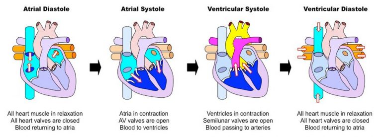
Fig. 8. Cardiac cycle

Ventricular Systole includes contraction of the left and right ventricles which is followed by electrical stimulation. During this AV valves are closed and semilunar valves are open. It takes about 0.3 seconds.

Cardiac diastole is a period when heart relaxes to fill the blood. When atria and ventricles are relaxing together, they form complete cardiac diastole. During ventricular diastole, pressure in the ventricles drops below the left atrial pressure, mitral valve opens and left ventricle gets filled with the blood. Similarly, when the pressure in the right ventricle drops below that in the right atrium, the tricuspid valve opens, and the right ventricle gets filled with blood. During diastole, the pressure within the left ventricle is lower than that in aorta, which allows the blood to circulate in the heart itself with the help of the coronary arteries.

Heart Sound

The heart sound is known as “lubb-dubb” sound. The first heart sound lubb is produced when mitral and tricuspid valves gets closed at the beginning of the ventricular systole. The second sound dubb is produced when aortic and pulmonary valves gets closed at the end of ventricular systole.
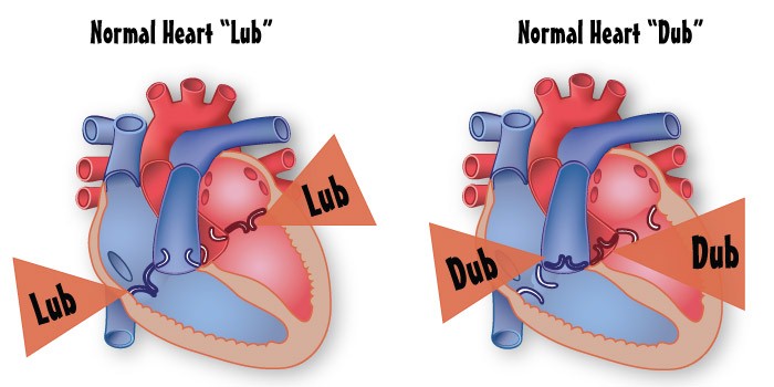
Fig. 9. Heart sound

Tuesday, 25 January 2022

Life Cycle of Mosquito-A study


Objective

Our objective is to study the life cycle of a mosquito.

The Theory

The mosquitoes are a family of small, midge-like flies. Like all flies, mosquitoes go through four stages in their life - egg, larva, pupa, and adult. We call this as the life cycle.  Each of these stages is morphologically different from the other, with even the habitat of each stage differing. The first three stages - egg, larva and pupa are largely aquatic, whereas the adult stage is aerial.

Mosquito Life Cycle

We will now look at the four distinct stages of development in the life cycle of a mosquito.

Stage 1 - Egg

The eggs are laid one at a time and they float on the surface of the water. Normally the eggs are white when first deposited, then darken to near black within a day. They hatch in one to three days depending on the temperature. Eggs left on moist soil can last for up to a year, until the ground is flooded again, before hatching.

In the case of Culex and Culiseta species, 200-300 eggs are stuck together in rafts. Anopheles and Aedes species do not make egg rafts but lay their eggs separately. Culex, Culiseta, and Anopheles lay their eggs on water while Aedes lay their eggs on damp mud. The eggs generally do not hatch until the place is flooded. Most eggs hatch into larvae within 48 hours. When the larvae are ready to hatch, they use a small temporary ‘tooth’ on their head to break open the egg along a suture that was made by it.

Stage 2 - Larva

Mosquito larvae, commonly called ‘wigglers’ or ‘wrigglers’, live in water from 7 to 14 days depending on the water temperature. Larvae swim either through propulsion with their mouth brushes, or by jerky movements of their entire bodies, giving them the common name of ‘wigglers’. The larva begins to feed on bacteria and decaying organic matter on the water surface, soon after they hatch out of eggs. They spend most of their time hanging upside down at the surface, sucking in oxygen through the siphon. The siphon is located at the base of their abdomen and is similar to a snorkel. Brushes that are located in front of their mouths collect the food. Anopheles larvae do not have a siphon and they lay parallel to the water surface. The larval stage lasts for a few days to a few weeks, during which the larvae shed several layers of their outer skin, called moulting. This allows further growth.

Stage 3 - Pupa

After the larvae have completed moulting, they become pupae. This is the stage in which they undergo metamorphosis to become an adult mosquito. The pupal stage is a resting, non-feeding stage. Mosquito pupae are commonly called ‘tumblers’. The pupa is lighter than water and therefore floats at the surface. The mosquito pupa is comma-shaped. The head and thorax are merged into a cephalothorax, with the abdomen curving around underneath. At one end of these curved bodies is the large head and at the other end is the flippers used for swimming. They must take in oxygen from time to time through two breathing tubes known as ‘trumpets’. After a few days or longer, depending on the temperature and other circumstances, the pupa rises to the water surface, the dorsal surface of its cephalothorax splits, and the adult mosquito emerges.

Stage 4 - Adult

The newly emerged adult rests on the surface of the water for a short time to allow itself to dry and harden its parts. Also, the wings have to spread out and dry properly before it can fly.

Adult mosquitoes have a head with two large compound eyes, a thorax, a pair of scaled wings and six jointed legs. They also have antennae and a proboscis. Adult mosquitoes mate within the first few days after emerging from the pupal stage.

It is the carbon dioxide that we exhale, and the lactic acid from our sweat that combine to make us smell like a mosquito buffet. Mosquitoes can pick up these smells from 100 feet, and they can also feel our body heat and notice movements.

Only female mosquitoes have the mouth parts necessary for sucking blood. When biting with their proboscis, they stab two tubes into the skin, one is an anti-coagulant to keep the blood flowing and is a mild painkiller that helps them escape detection, the other helps to suck blood. They use the blood not for their own nourishment but as a source of protein for their eggs. For food, both males and females eat nectar and other plant sugars.

Some interesting mosquito facts

  • There are over 2500 different species of mosquitoes.
  • The feeding habits of mosquitoes are quite unique in that it is only the adult females that feed on blood. The male mosquitoes feed only on plant juices.
  • Mosquitoes must have water in which to complete their life cycle.
  • Most female mosquitoes need to feed on animal blood before they can develop eggs.
  • A female can produce up to 500 eggs before she finally dies.
  • Mosquitoes don't travel more than a mile from the place where they were hatched.
  • The length of life of the adult mosquito usually depends on factors like - temperature, humidity, sex of the mosquito and time of the year.
  • Once mosquitoes emerge from their pupal cocoons and take flight, male mosquitoes last less than a week and the females’ maybe a couple of months.

Learning Outcomes

  1. Students understand the different stages of a Mosquito life cycle.
  2. Students get to know different types of Mosquitoes and the diseases spread by them.
  3. Students understand the differences in each stage of the mosquito life cycle through the animated demonstrations.

Binary Fission and Budding -A Study


Objective

Our objective is to study using the prepared slides;

  1. Binary fission in Amoeba.
  2. Budding in Yeast.

The Theory

Reproduction

All living things produce their own kind through the process called reproduction. Reproduction takes place sexually and asexually.

Sexual reproduction

Sexual reproduction is the primary method of reproduction for the vast majority of macroscopic organisms, including almost all animals and plants. Here are two main processes during sexual reproduction in eukaryotes: meiosis, involving the halving of the number of chromosomes; and fertilisation, involving the fusion of two gametes and the restoration of the original number of chromosomes. During meiosis, the chromosomes of each pair usually cross over to achieve homologous recombination that helps produce genetic diversity when cells divide in meiosis.

Asexual reproduction

Asexual reproduction is the primary form of reproduction for single-celled organisms such as the archaea, bacteria, and protists. Many plants and fungi reproduce asexually as well. Asexual reproduction is a mode of reproduction by which offspring arise from a single parent, and inherit the genes of that parent only. The offspring will be the exact genetic copies of the parent.

New organisms are produced in rapid multiplication by the process of amitotic or mitotic divisions. Amitosis is the process by which a cell directly separates, as the nucleus and cytoplasm are directly cut in two. Mitosis is the process by which a cell, which has previously replicated each of its chromosomes, separates the chromosomes in its cell nucleus into two identical sets of chromosomes, each set will have its own new nucleus. It is a form of nuclear division.

Binary fission and budding are two common method of asexual reproduction. Binary fission is found in unicellular organisms like Amoeba, Paramaecium and Euglena, to name and few. Budding is found in Yeast and Hydra.

Binary fission in Amoeba

Amoeba is a shapeless tiny unicellular organism that has a porous cell membrane which encloses the cell organelles and cytoplasm. Amoeba reproduces by the common asexual reproduction method called binary fission. After replicating its genetic material through mitotic division, the cell divides into two equal sized daughter cells. The genetic material is also equally partitioned; therefore the daughter cells are genetically identical to each other and the parent cell. In this process, the nucleus of the Amoeba first divides to form two daughter nuclei by the process of Karyokinesis. After the nucleus has divided into two, the process of Cytokinesis takes place in which the cytoplasm in the mother cell divides into two daughter cells. This leads to the formation of the two daughter Amoebae cell having a nucleus and its own cell organelles.

Karyokinesis is the process of the division of the nucleus. It corresponds to the separation of the daughter chromosomes into two daughter nuclei. Karyokinesis is usually followed by Cytokinesis.

Cytokinesis is the process of the division of the cytoplasm. It corresponds to the separation of the daughter nuclei into two daughter cells. Cytokinesis occurs immediately after mitosis.


Budding in Yeast

Yeast are unicellular (some are multicellular) eukaryotic micro-organisms belonging to the kingdom fungi. Yeast size can vary greatly depending on the species, typically measuring 3-4 µm in diameter. Most yeasts reproduce asexually by an asymmetric division process called budding. First it produces a small protuberance on the parent cell that grows to a full size and forms a bud. The nucleus of the parent cell splits into a daughter nucleus and migrates into the daughter cell. The bud detaches from the mother’s body by forming a constriction at the base. Budding will repeat to form a chain of bud cells. The daughter cell produced during the budding process is generally smaller than the mother cell.


Learning Outcomes

  1. Students understand the terms budding, binary fission and a few other terms.
  2. Students understand the different stages of binary fission in Amoeba.
  3. Students understand the different stages of budding in Yeast.
  4. Students understand the experiment better through the animated demonstration.

Wednesday, 1 September 2021

To experimentally demonstrate that carbon dioxide is released during the process of respiration.


Aim

To experimentally demonstrate that carbon dioxide is released during the process of respiration.

Principle/Theory

The process of respiration is biochemically carried out wherein food, glucose to be precise is oxidized and energy is released. In this experiment, gram seeds (moistened) are used. The purpose of using these seeds is that they release carbon dioxide and are respiring actively. The released carbon dioxide is consumed by the solution of KOH.

Material Required

  • Soaked gram seeds
  • U-shaped delivery tube
  • Conical flask
  • Blotting paper (moist) /cotton wool
  • Thread
  • Water
  • Beaker
  • Test tube
  • Rubber cork with a single hole
  • Freshly prepared KOH solution (20%)
  • Vaseline

Procedure

  • Germinate close to 25 seeds. This can be done by wrapping them in moist blotting paper or cotton wool for around 3 to 4 days
  • Set up the germinated or sprouted seeds in the conical flask. Spray some water into the flask to dampen the seeds
  • With the help of a thread, suspend the conical flask containing the test tube having a freshly prepared 20% KOH solution.
  • Use the rubber cork to seal the opening of the conical flask.
  • One edge of the U-shaped glass delivery tube present in the conical flask should be inserted through the hole in the rubber cork. The other edge should be placed into a beaker that is saturated with water
  • All attachments of the set-up should be sealed. This can be done using vaseline to create an air-tight environment
  • The initial water level present in the U-shaped delivery tube needs to be marked.
  • Leave the experimental set-up uninterrupted for 1 to 2 hours. Observe the fluctuations in the water level in the tube.

Observation

Careful observation after a certain period of time reveals that the water level in the U-shaped delivery tube has risen in the beaker.


Conclusions

The rise in level water indicates that carbon dioxide is released as a result of germinating gram seeds during the process of respiration in the conical flask. The carbon dioxide that is released in the process is absorbed or consumed by the KOH solution that is suspended in the test tube in the conical flask, creating a vacuum or a void in the flask resulting in the upward water movement in the tube. Hence, the water level in the tube changes.

Precautions

  • The seeds that are to be germinated needs to be moistened
  • Air-tight environment for all the connections in the experimental set-up
  • The KOH solution that is used needs to be freshly prepared
  • Care needs to be taken to ensure that one end of the delivery tube is placed in the conical flask. The other edge is submerged in the water of the beaker
  • The tube that contains the KOH solution needs to be suspended carefully

Friday, 22 January 2021

Detection of Albumin in Urine


Sulphosalicylic Acid Test

Procedure

  • Take 2 ml urine sample in a measuring cylinder from the urine sample bottle.
  • Take a test tube and pour the urine in the test tube.
  • Using a dropper, take some sulphosalicylic acid.
  • Add few drops of sulphosalicylic acid in the test tube containing urine. A whitish color appears in the solution.
  • Using a test tube holder, hold the test tube firmly and heat it gently upon a burner.
  • A whitish or cloudy turbid solution indicates the presence of albumin in the urine sample.

Study of distribution of stomata


 Lab Procedure

  • Pluck one fresh leaf of a four-o’clock plant.
  • Take two watch glasses and pour some distilled water into the both watch glasses.
  • Split the leaf from the four-o’clock plant obliquely.
  • Take the peel from the upper surface of the leaf using the forceps.
  • Place the peel into a watch glass containing water.
  • Take another peel from the lower surface of the leaf using the forceps.
  • Place the peel into the other watch glass containing water.
  • Using a dropper, take few drops of Safranin solution and put it into the two watch glasses.
  • Take two clean glass slides and place the leaf peel on the slides one by one, using a brush.
  • Take a blade and cut a small rectangle or square piece from each peel.
  • Take some glycerine using a dropper and put one drop of glycerine on both slides.
  • Take a cover slip and place it gently on the peel with the help of a needle.
  • Take the glass slide and place it under compound microscope.
  • Observe under the microscope.
  • Count the number of stomata in the peels of both upper and lower epidermis of the leaf appearing in the microscopic field.

Observations

  • The number of stomata is greater in the lower epidermis, and fewer are present in the upper epidermis of the leaf taken from a four-o’clock plant.

Precautions

  • The curling of the peel should be avoided.
  • Always use a brush to transfer the peel from watch glass to the slide

XI BIO LAB WORK-Pigment separation by using paper chromatography


 Procedure

  • Take a few freshly plucked green spinach leaves.

  • Using scissors,  cut the spinach leaves into small pieces and let them fall into the mortar.

  • Take a measuring cylinder that contains 5ml of acetone and pour it into the mortar.

  • Grind the spinach leaves using the mortar and pestle.

  • Place the extract into a watch glass using a spatula.

  • Take a strip of filter paper having a narrow notch at one end of the strip.

  • Take a pencil and a scale and draw a horizontal line with a pencil about 2-3 cm away from the tip of the notch.

  • Put a drop of the pigment extract in the middle of the line with the help of a capillary tube.

  • Allow the drop to dry and repeat till four or five drops are placed on the paper.

  • Take the chromatographic chamber and pour ether acetone solvent in it.

  • Fold one end of the filter paper strip and staple it.

  • Using a thread, hang the filter paper strip in the chromatographic chamber.

  • The loading spot should remain about 1 cm above the solvent level.

  • Leave the chromatographic chamber undisturbed for some time.

  • We can observe, as the solvent moves through the paper, it spreads the different pigments of the mixture to various distances.

  • When the solvent rises about 3/4th up the strip, remove the strip carefully and let it dry.

XI Biology lab work- to test sugar in urine.


1.Benedict's Test

Materials required:

Test tube, test tube holder, urine sample, measuring cylinders, Benedict’s solution and burner.

Procedure:

  • Take 2 ml urine sample in a measuring cylinder from the urine sample bottle.
  • Take a test tube and pour the urine sample in it.
  • Take 5 ml Benedict’s reagent in a measuring cylinder.
  • Add Benedict’s reagent to the test tube that contains urine sample.
  • Using a test tube holder, hold the test tube firmly and heat it for 2 minutes on the burner.
  • Keep shaking the test tube while heating.
  • A yellow precipitate appears which indicates the presence of sugar in urine.
  • Depending upon the concentration of sugar in the urine, either green, yellow, or brick red precipitates are formed.

2.Fehling's test

Materials required

Test tube, test tube holder, urine sample, measuring cylinders, Fehling’s solution A, Fehling’s solution B and burner.

Procedure

  • Take 2 ml urine sample in a measuring cylinder from the urine sample bottle.
  • Take a test tube and pour the urine sample in it.
  • Take 2 ml Fehling’s solution A in a measuring cylinder.
  • Add Fehling’s solution A to the test tube that contains urine sample.
  • Take 2 ml Fehling’s solution B in a measuring cylinder.
  • Add Fehling’s solution B to the test tube that contains urine sample.
  • Using a test tube holder, hold the test tube firmly and heat it gently for 2 minutes on the burner.
  • Keep shaking the test tube while heating.
  • A green precipitate appears which indicates the presence of traces of sugar in urine.
  • Depending upon the concentration of sugar in the urine, either green, yellow or brick red precipitates are formed.

Sunday, 20 December 2020

Endocrine System 2


Pancreas

Pancreas is a composite gland which acts as both exocrine and endocrine gland. 

The endocrine' pancreas consists of 'Islets of Langerhans'. There are about 1 to 2 million Islets of Langerhans in a normal human pancreas representing only 1 to 2 per cent of the pancreatic tissue. The two main types of cells in the Islet of Langerhans are called a-cells and b-cells. The a-­cells secrete a hormone called glucagon, while the b-cells secrete insulin.

Glucagon is a peptide hormone, and plays an important role in maintaining the normal blood glucose levels. Glucagon acts mainly on the liver cells (hepatocytes) and stimulates glycogenolysis resulting ill an increased blood sugar (hyperglycemia).

T.S. of Pancreas

Glucagon reduces the cellular glucose uptake and utilisation, Thus, glucagon is a hyperglycemic hormone.

Insulin is a peptide hormone, which plays a major role in the regulation of glucose homeostasis. Insulin acts mainly on hepatocytes and adipocytes (cells of adipose tissue), and enhances cellular glucose uptake and utilisation.

Insulin also stimulates conversion of glucose to glycogen (glycogenesis) in the target cells.

Prolonged hyperglycemia leads to a complex disorder called diabetes mellitus which is associated with loss of glucose through urine and formation of harmful compounds known as ketone bodies.


Testis

A pair of testis is present in the scrotal sac (outside abdomen) of male individuals.

Testis performs dual functions as a primary sex organ as well as an endocrine gland.

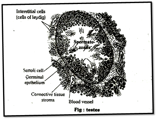
Testis is composed of seminiferous tubules and stromal or interstitial tissue. The Leydig cells or interstitial cells, which are present in the intertubular spaces produce a group of hormones called androgens mainly testosterone. 

Testes

Androgens regulate the development, maturatior and functions of the male accessory sex organs like epididymis, vas deferens, seminal vesicles, prostate gland, urethra etc. These hormones stimulate muscular growth, growth of facial and axillary hair, aggressiveness, low pitch of voice etc. Androgeru playa major stimulatory role in the process of spermatogenesis (formation of spermatozoa), Androgens act on the central neural system and influence the male sexual behaviour (libido). These hormones produce anabolic (synthetic) effects , on protein and carbohydrate metabolism. 
 

Ovary

Females have a pair of ovaries located in the abdomen.

Ovary is the primary female sex organ which produces one ovum during each menstrual cycle. In addition, ovary also produces two groups of steroid hormones called estrogen and progesterone. 

Ovary

Ovary is composed of ovarian folliclos and stromal tissues.

The estrogen is synthesised and secreted mainly by the growing ovarian follicles, After ovulation, the ruptured follicle is converted to a structure called corpus luteum, which secretes mainly progesterone.

Estrogens produce wide ranging actions such as stimulation of growth and activities of female secondary 'sex organs, development of growing ovarian follicles, appearance of female secondary sex characters (e.g., high pitch of voice, etc.), mammary gland development.

Progesterone supports pregnancy. Progesterone also acts on the mammary glands and stimulates the formation of alveoli (sac-like structures which store milk) and milk secretion.
 

Mechanism of Hormone Action

Hormones produce their effects on target tissues by binding to specific proteins called hormone receptors located, in the target tissues only.

Each receptor is specific to one hormone only and hence receptors are specific.

Hormone-Receptor complex formation leads to, certain biochemical changes in the target tissue, 'Target tissue metabolism and hence physiological functions are regulated by hormones.

The hormone receptor complex may acts in one or the two ways –

Formation of cAMP: Mechanism of formation of cAMP was discovered by E.W. Sutherland in 1950. The hormone receptor complex causes the release of an enzyme adenyl cyclase, from the receptor site. This enzyme hydrolyses the A. TP into c-AMP. The c-AMP activates the existing enzvmo system of the cell. This accelerates the metabolu­reactions in cell. The hormone is called firs: messenger and the c-AMP is termed the second messenger. e.g., Adrenaline causes the secrertion of glucose from the liver cell from this mechanism. 

Mechanism of Hormone Action on Cell Surface

(ii) Change in membrane permeability: The receptor proteins of some hormones are large transmembrane intrinsic protein acting as ion channels for facilitated diffusion of Na+, K+, Ca2+ etc. On binding with specific hormone these receptor proteins undergo conformational changes, so that the membrane permeability for ions is altered, resulting into important changes in metabolism.

For example, insulin promotes the entry of glucose from blood into the muscles cells by increasing the permeability of- sarcolemma to glucose.

The steroid hormones act within the cell. Their small, lipid soluble molecules pass through the cell membrane and bind to specific receptor molecules present in the cytoplasm. The receptor molecules carry them into the nucleus. Here, the receptor hormone complex binds to a specific receptor site on the chromosome and activates certain genes that were previously repressed. The activated gene transcribe m-RNA which directs the synthesis of enzyme (protein molecule) in the cytoplasm. The enzyme molecule promote the metabolic reactions in the cell. 

Mechanism of Cell Surface Within a Cell
Note:

The atrial wall of our heart secretes a very important peptide hormone called atrial natriuretic factor (ANF), which decreases blood pressure. When blood pressure is increased, ANF is secreted which causes dilation of the blood vessels. This reduces the blood pressure.

The juxtaglomerular cells of kidney produce a peptide hormone called erythropoietin which stimulates erythropoiesis (formation of RBC).

Endocrine cells present in different parts of the gastro-intestinal tract secrete four major peptide hormones, namely gastrin, secretin, cholecystokinin (CCK) and gastric inhibitory peptide (GIP).

Gastrin acts on the gastric glands and stimulates the secretion of hydrochloric acid and pepsinogen. Secretin acts on the exocrine pancreas and stimulates secretion of water and bicarbonate ions.

CCK acts on both pancreas and. gall bladder and stimulates the secretion of pancreatic enzymes and bile juice, respectively. GIP inhibits gastric secretion and motility.

Hormones which interact with intracellular receptors (e.g., steroid hormones, iodothyronines, etc.) mostly regulate gene expression or chromosome function by the interaction of hormone-receptor complex with the genome.

Heterocrine gland: These are those endocrine glands which are involved in hormone secretion as well as some other function eg. pancrease, gonads, placenta, GI mucosa and kidneys.

In females prolactin induce maternalism i.e. strong emotional attachment.

In male prolactin promotes paternalism i.e. protective attitudes towards family members and intensive food gathering for the family.

Contrary to thyroid dwarf (cretins), the pituitary dwarf have a normal mental development and proportionate body.

Sporadic cases of simple goitre (sporadic goitre) in a population are normally due to genetic defect.

Muller organ and subneural gland is homologous to pituitary gland.

Tropic hormone: A hormone which stimulates another endocrine gland to secrete its hormone is called trophic hormone.

Simmond's disease: This condition is due to atrophy of the anterior lobe of pituitary gland.

Pheochromocytoma: It is due to hypersecretion of adrenaline causes, high blood pressure, high level of sugar in blood and urine, high metabolic rate, nervousness and sweating.

Sexual pseudoprecocity results from adrenal cortex, testes, ovary or from other sources, including extragonadial tumours.

Eunuchoidism: Failure of testosterone secretion in male-causes eunuchoidism.

eunuch has a undeveloped secondary sex organs like prostrate, seminal vesicle and penis

does not produce sperm.

Growth hormone: Stimulate the liver to form "Somatomedins" ("Insulin like growth factors"). This somatomedins potent effect to bone growth.

In heart cGMP has antagonistic effect to cAMP, cAMP mediate muscle contraction in response to adrenaline, while cGMP slow down muscle contraction in response to acetylcholine.

cGMP is used as second messenger in atrial natriuretic peptide and nitric oxide.
 

Difference between Nervous and Endocrine Coordination


Nervous Co-ordination


Endocrine coordination

1

Information passes as electrical impulses along nerve fibres.

1

Information passes as a chemical substance through the blood and lymph.

2

There is rapid transmission of information.

2

There is slow transmission of information.

3

Response is immediate.

3

Response is usually slow,

4

Response is very exact.

4

Response is usually widespread.

5

Response is short lived.

5

Response is long-lasting.


Thyroid gland is the largest endocrine gland in the body.

Endostyle of lower vertebrates like Herdmania, Amphioxus is homologous of thyroid gland.

Thyroid is the only endocrine gland in the body which stores its hormone in its inactive state.

The oxidation of iodine is promoted by the enzyme peroxidase.

21 October is Iodine deficiency day

Thyroxine stimulates the metamorphosis of tadpole larva in amphibians.
 

Difference between Hormone and Enzymes

S.No.

Characters

Enzymes

Hormones

1.

Chemistry

Always proteinaceous

May be proteinaceous, or amine or steroids.

2.

Molecular weight

Macromolecules with high molecular weights.

Have low molecular weights.

3.

Diffusibility

Non-diffusible through cell membrane.

Diffusible through cell membrane.

4.

Site of action

Either act intracellalarly or carried by some duct to another site.

Generally carried by blood to a target organ.

5.

Mode of action

Always act as biocatalysts and increase the rate of metabolic physiological process.

May be excitatory or inhibitatory in their physiological action.

6.

Reversibility

These catalyze reversible reactions.

Hormone controlled reactions are not reversible.

7.

Effect of concentration

Reaction rate increase with increase in their concentration upto a limit.

Deficiency or excess of hormone causes metabolic disorders and diseases,

8.

Speed

Act quickly

Some are quick acting, while some are slow acting with a lag period.

9.

Consumption

Not used in metabolic functions.

Used up in metabolic functions.


Number of Hormones Secreted by Different Endocrine Glands

Endocine-glands

Number of secreted hormones

Pituitary - Anterior

-

7

Hypothalamus

-

2

Pineal body

-

2

Thymus

-

3

Thyroid

-

2

Parathyroid

-

1

Islets of Langerhans

-

3

Adrenal cortex

-

46

Adrenal medulla

-

2

Testes

-

1

Ovary

-

3

Placenta

-

2

Kidneys

-

2

Stomach

-

1

Duodenum

-

5

Ileum

-

2


Anterior Pituitary Hormones

Hormone and Target Tissues

Principal Action

Hormone and Target Tissues

Principal Action

Human growth hormone (hGH) or somatotropin

Stimulates liver, muscle, cartilage, bone, and other tissues to synthesize and secrete insulin like growth factor of body cells, protein synthesis tissue repair, glucose concentration.

Prolactin (PRL)

Together with other hormones, promotes milk secretion by the mammary glands.

Thyroid -stimulating hormone (TSH) or thyrotropin

Stimulates synthesis and secretion of thyroid hormones by thyroid gland.

Adrenocorticotropic hormone (ACTH) or corticotropin

Stimulates secretion of glucocorticoids (mainly cortisol) by adrenal cortex.

Follicie- stimulating hormone (FSH)

In females, initiates development of occytes and induces ovarian secretion of estrogens. In males stimulates testes to produce sperm,

Melanocyte-stimulating hormone (MSH)

Exact role in humans is unknown but may influence brain activity, when present in excess, can cause darkening of ski


Posterior Pituitary Hormones

Hormone and Target Tissues

Control of Secretion

Principal Actions

Oxytocin (OT)

Neurosecretory cells of hypothalamus secrete OT in response to uterine distention and stimulation of nipples.

Stimulates contraction of smooth muscle cells of uterus to cause child birth; stimulates contraction of myoepithelial cells in mammary glands to cause milk ejection.

Antidiuretic hormone (ADH) or vasopressin

Neurosecretory cells of hypothalamus secrete ADH in response to elevated blood osmotic pressure, dehydration loss of blood volume. Pain or stress; low blood osmotic pressure, high blood volume, and alcohol to inhibit ADH secretion.

Conserves body water by, decreasing urine volume; decreases water loss through perspiration: raises blood pressure by constricting arterioles


Hormones, Their Chemical Nature and Functions

S.No.

Name of endocrine gland

Name of hormone and its chemical nature

Functions

1.

Neurosecretory     cells of Hypothalamus     (Supraoptic Nucleus and Paraventricular Nucleus)

Oxytocin and vasopressin monopeptide Gonadotropin releasing hormones Other releasing hormones e.g. TSHRH, MSHRH, ACTHRH, GHRH etc. Proteinaceous

Milk ejection and parturition (oxytocic effect). Vasoconstriction and antidiuretic (vasotocin) effects. Stimulates FSH and LH synthesis Stimulate TSH, MSH, ACTH GH secretions from pituitary.

2

Pituitary (a) Neurohypophysis (Pars Nervosa) (b) Adenohypophysis contains diverse cell types)

Store and release Oxytocin and Vasopressin. Proteinaceous or glycoprotein

Hormone release is related to physiological state and requirements. Affect growth, development differential pubertal changes and other metabolic mechanism

3.

Pineal

Melatonin-derived from the amino acid tyrosine

Antagonist to FSH / LH   Regulates biological/circadian rhythms.

4.

Thyroid gland (amine hormone) having - NH2 group)

(a) Thyroxine, iodinated amino acid called tyrosine (T2, T3, T4). (b) Thyrocalciton in (Peptide)

(a) Controls basal metabolic rate (BMR). All organ / system of body responds to thyroxine. (b) Facilitates Ca+2 absorption

5.

Parathyroid gland

Parathormone, Peptide

Ca+2 and PO-4 metabolism.

6.

Thymus

Thymosine (polyneotide)

Anti-FSH and LH; dela; delays puberty

7.

Islets of lengerhans


(i) Gluconeogenesis / Glycogenolysis (ii) G lycogenesis (iii) Gastric functions    

8.

(= Endocrine pancrease) (i) a-cells (ii) b-cells (iii) d-cells

(a) Catecholamines (epinephrine = adrenaline, and norepinephrine = noradrenaline (derived from tyrosine) (b) Mineralcorticoids and glucocorticoids and traces of androgen and estrogen steroids derived from cholesterol

Stresses = emergency = Fright, Fight and Flight Hormone (3F) acclerates cardiac functions muscle activity etc. (b) Electrolyte and carbohydrate metabolism.

9.

Adrenal gland (a) Adrenal medulla (Amine hormone have – NH2) (b) Adrenal cortex

Estrogen (Steroid) Estrone, estradiol Estrogen and Progesterone (Steroid) (a)Steroid secreted are estrogen and progesterone (b) Relaxin-Polypeptide

(a) Secondary sex character primary action on uterine endometrium mitogenic. (a) Secreted during -luetal phase of menstrual cycle in human female and oestrous cycle of other mammals. Prepares uterine endometrium for receiving blastocytes for implantation, Progesterone is also called pregnancy hormone and is anti-FSH and anti-LH/anti-LTH. (a) Maintenance of pregnant state, prevents lactogenesis folliculogenesis, and Ovulation. (b) Act on pubic symphysis and enlarges the birth canal to facilitate birth. Acts synergestically with oxytocin during this process (parturition)

10.

Ovary (a) Granulosa cells steroid fat soluble have sterol group derived from cholesterol (b) Corpus luteum Placenta temporary endocrine gland formed during pregnancy 

Inhibin - Polypeptide (ii) Estradiol-Steroid Androgens (e.g Testosterone) Steroid androstenedione)

Inhibits FHS action and attenuates spermatogenesis decrementally -do- (i) Pubertal changes in male, (ii) Seco. Sex characters in male, (iii) Sex drives. (iv) Spermatogenesis

11.

Testis (i) Sertoli cells (= sustentacular cells) (ii) Leydig cells (=Interstitial cells)

Gastrin (i) Secretin (ii) Cholecystokinin (CCK) (iii) Enterogastrone (iv) Duocrinin, (v) Enterokinin     (vi) Villikrinin

Stimulates gastric juices secretion from gastric gland, movement of sphincters of stomach and increased movement of stomach (i) Stimulates secretion of succus entericus, (ii) Bile released from gall bladder, (iii) Inhibits gastric secretin, (iv)  Stimulates secretion of mucous from Brunner's gland, (v) Stimulate intestinal gland, (vi) Stimulate villi movement.


Disease Caused by Hormonal Irregularities

Disease

Hormone

Quantity

Gland

Dwarfism

GH

Deficiency

Pituitary

Gigantism

GH

Excess

Pituitary

Acromegaly

GH

Excess

Pituitary

Simmond's disease

GH

Deficiency

Pituitary

Diabetes insipidus

ADH

Deficiency

Pituitary

Cretinism

Thyroxine

Deficiency

Thyroid

Simple goitre

Thyroxine

Deficiency

Thyroid

Myxaedema

Thyroxine

Deficiency

Thyroid

Exophthalamic goitre

Thyroxine

Excess

Thyroid

Tetani

Parathyroid

Deficiency

Parathyroid

Plummer's disease

Thyroxine

Excess

Thyroid

Addison's disease

Mineralocorticoids (Aldosterone)     and Glucocorticoids (cortisol)

Deficiency

Adrenal cortex

Conn's 'disease

Mineralocorticoids

Excess

Adrenal cortex

Cushing's disease

Corticosteroid

Excess

Adrenal cortex

.

MCQ MOLECULAR BASIS OF INHERITANCE

  1. Amino acid sequence, in protein synthesis is decided by the sequence of ( a ) r RNA ( b ) t RNA ( c ) m RNA ( d ) c DNA 2. Antipa...